Fig. 1.
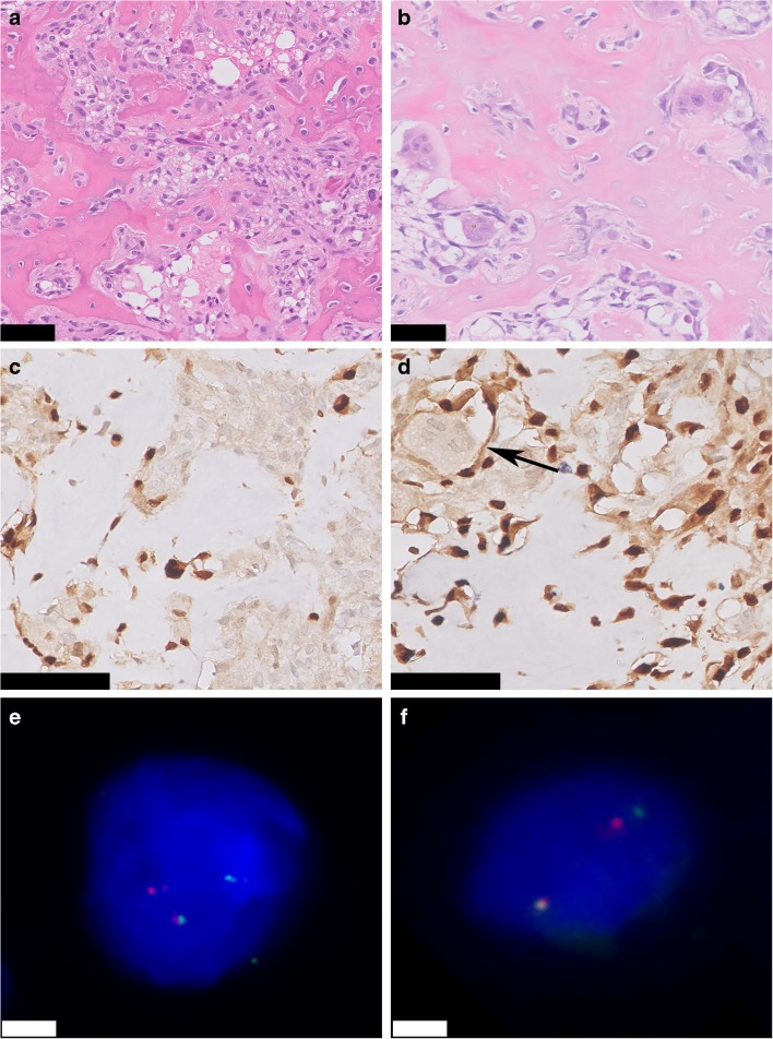
Osteoid osteoma and osteoblastoma. Hematoxylin & Eosin (H&E) staining of the nidus of osteoid osteoma a and osteoblastoma b. Regular deposited trabeculae of woven bone are surrounded by active osteoblasts. Both osteoid osteoma and osteoblastoma show strong and diffuse nuclear expression of FOS. Osteoclast-like giant cells are negative (arrow) c and d. Fluorescence in situ hybridization (FISH) using split-apart probes for FOS shows a segregated red and green signal in both osteoid osteoma and osteoblastoma, indicating a FOS rearrangement e and f. Scale bar, 50 μm a–d. Scale bar, 5 μm e and f

