Fig. 3.
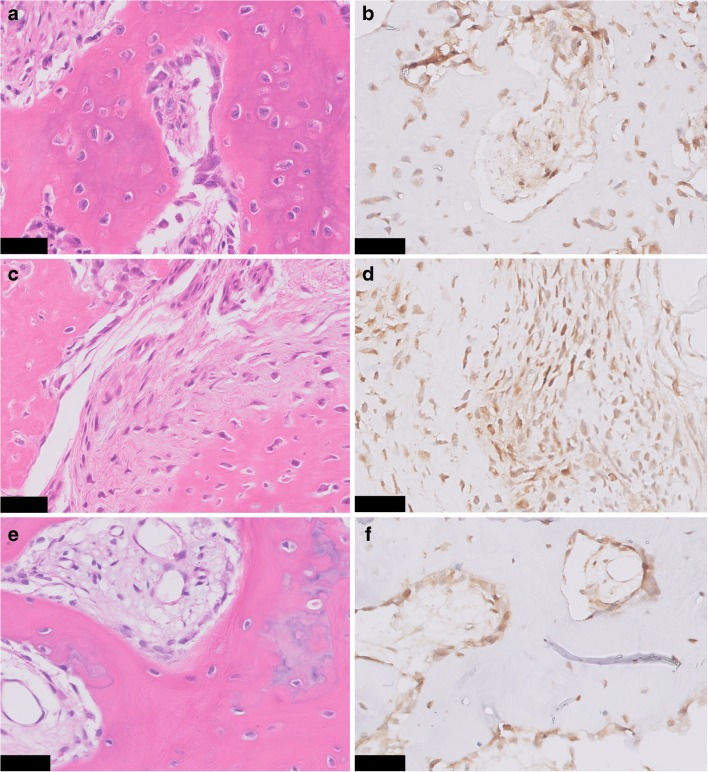
FOS immunohistochemistry in proliferative bone lesions. Myositis ossificans, with peripheral zone showing ill-defined trabeculae of woven bone, rimmed with osteoblasts (H&E) a. Immunohistochemistry of FOS showing moderate nuclear staining of osteoblasts b. Bizarre parosteal osteochondromatous proliferation with a disorganized mix of woven bone and spindle cells (H&E) c, where additional FOS immunohistochemistry shows moderate staining in both components d. Central area with trabecular bone in subungual exostosis (H&E) e, showing moderate expression of FOS in osteoblasts f. Scale bar, 50 μm (a–f)

