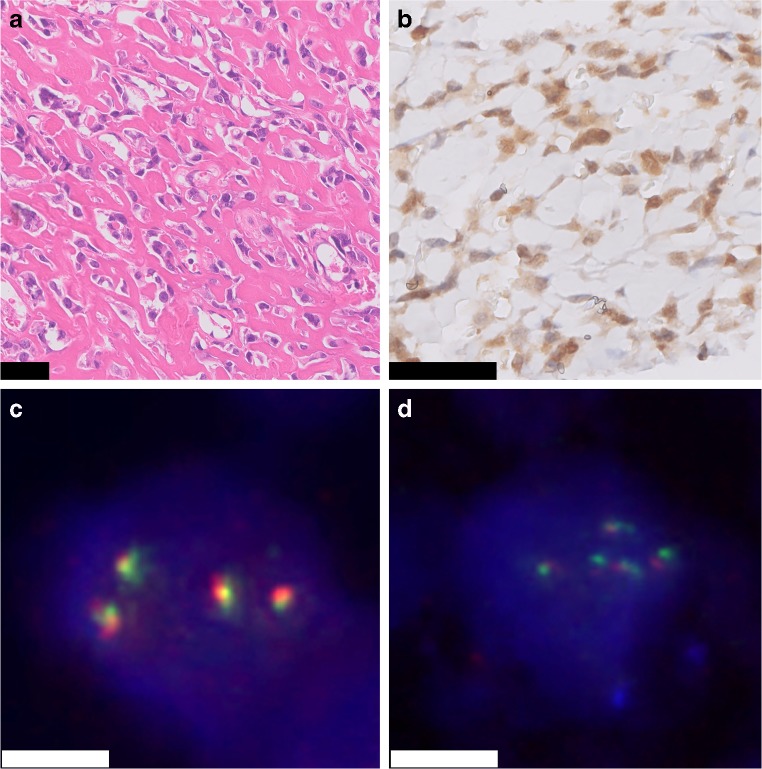
Fig. 4.
High-grade osteoblastic osteosarcoma. H&E staining shows atypical tumor cells depositing lace-like osteoid (H&E) (A). Immunohistochemistry for FOS shows moderate to strong nuclear staining of the tumor cells (B). Additional FISH for FOS and FOSB shows gains of the FOS- and FOSB-locus, respectively (C and D). Scale bar, 50 μm (A and B). Scale bar, 5 μm (C and D)

