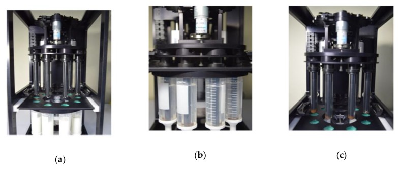
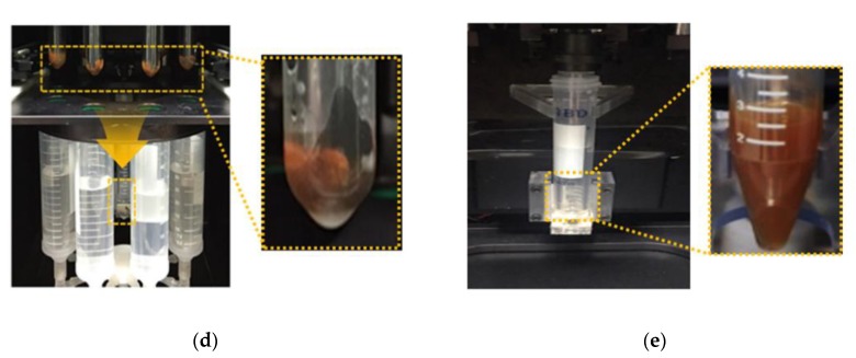
Figure 2.
Representative step-by-step images of the automated IMS process: (a) Introduction of a pre-enriched sample containing immunomagnetic beads bound with target bacteria in a milk sample; (b) Separation of immunomagnetic beads from the sample solution using an inserted magnetic bar; (c) Removal of the adhered magnetic beads along with the target bacteria from the sample solution; (d,e) Immersion of each glass cylinder in to the recovery tube containing 2 mL of buffer solution and redispersion of the magnetic beads and bacteria by vertical movement (two times repetition).


