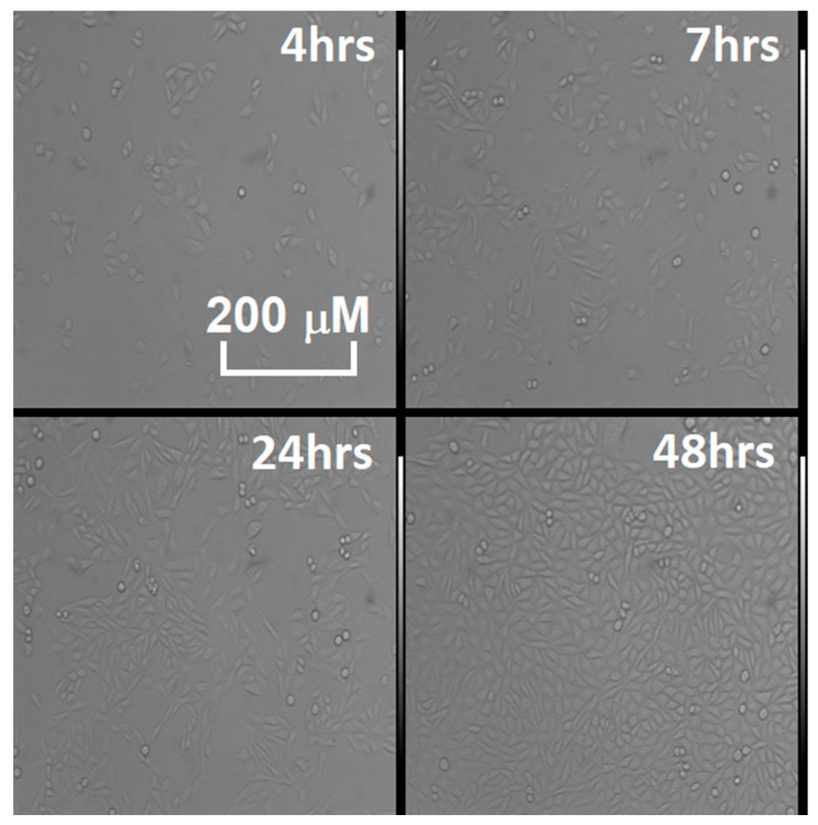
Figure 3.
Time lapse of the proliferation in the various CHO cell cultures tested in Figure 2, observed through the optical microscope (model: TE 300, Nikon magnification 10X). The living, plated cells are the elongated semitransparent corpuscles, while the dead ones are the round shapes, floating in the breeding ground.

