
Fig. 1.
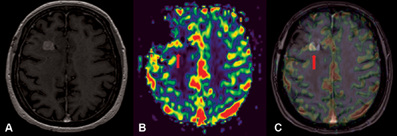
Advanced magnetic resonance imaging of the case. ( A ) Contrasting axial image in T1 with gadolinium, ( B ) perfusion map rCBV, ( C ) image fusion rCBV/MRI. Nodular area of contrast enhancement in the dorsal margin of the biopsy tract, with elevated rCBV, suggesting hyperperfusion (arrows). MRI, magnetic resonance imaging; rCBV, relative cerebral blood volume.