
Fig. 2.
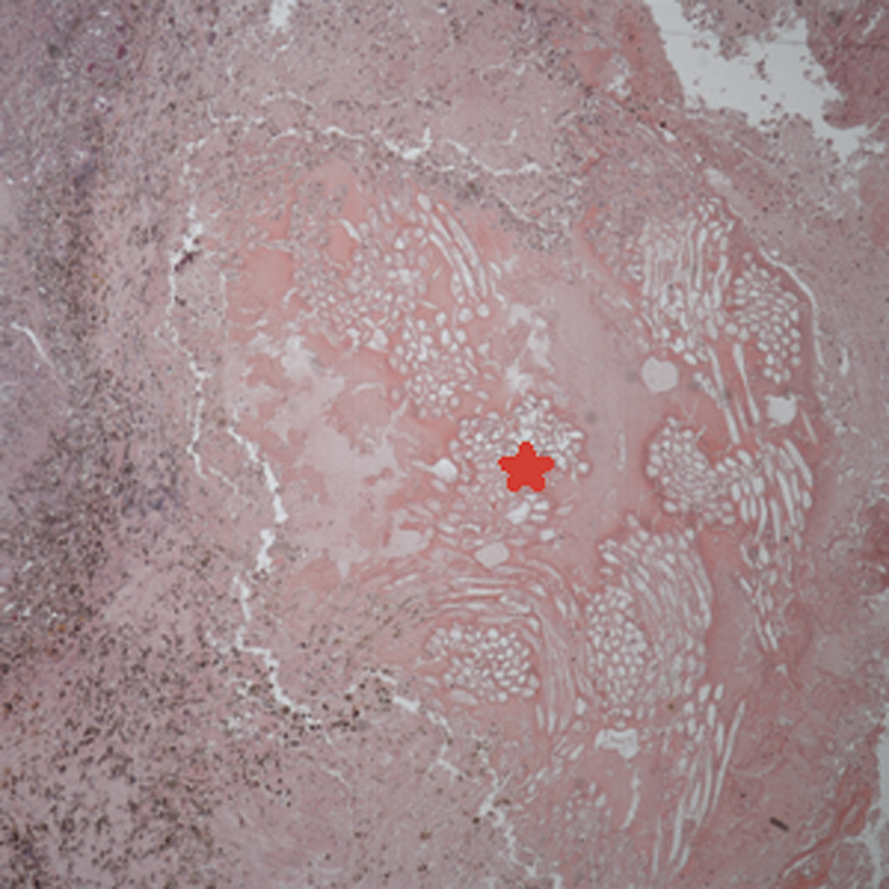
Low power (×40 original magnification) micrograph of Txm showing optically clear bundles of foreign material ( * ) with morphology consistent with oxidized cellulose that is used for hemostasis. The left portion of the micrograph shows fibrosis, hemosiderin-laden macrophages, and chronic inflammation (H & E). H & E, hematoxylin and eosin.