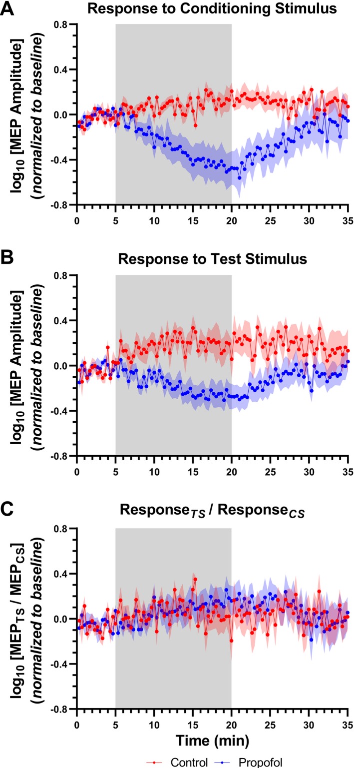
Figure 2.

Dose‐dependent decrease in MEP amplitude after propofol bolus. Data are presented as mean ± SEM of MEP amplitude recorded at baseline and at follow‐up timepoints. Following a short baseline, animals received an increased infusion rate of 2 mg/kg per min during 15 min, before returning to 1 mg/kg per min for another 15 min. A control group received a continuous infusion rate of 1 mg/kg per min for the entire 30‐min period. The shaded region indicates the timing of propofol infusion rate changes. (A, B) Increasing the infusion rate from 1 to 2 mg/kg per min resulted in a progressive decrease in the MEP amplitudes in response to the conditioning and test stimuli, respectively. When the infusion rate was restored to 1 mg/kg per min, a progressive increase in both responses was observed. (C) No changes in the inhibition ratio were observed.
