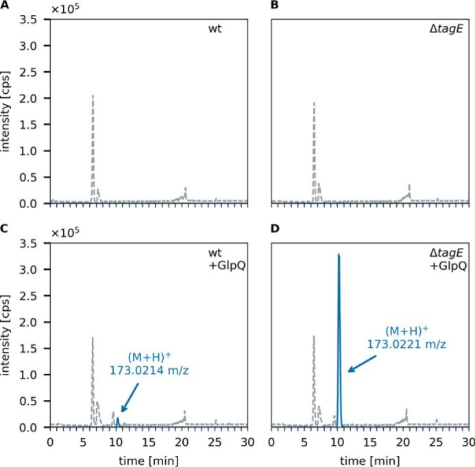
Figure 4.

GlpQ predominantly releases Gro3P from the cell walls of ΔtagE B. subtilis 168. The purified cell wall of B. subtilis (containing peptidoglycan and covalently bound WTA (PGN-WTA complex)) was incubated with GlpQ, and the formation of reaction products was analyzed by LC-MS. Shown are the BPC for mass range (M+H)+ = 120–800 (gray dashed lines) and the EICs of glycerolphosphate (M+H)+ m/z = 173.022 ± 0.02 (blue solid lines). A and B, analysis of the WT containing partially glycosylated WTA and ΔtagE containing nonglycosylated WTA cell walls in the absence of GlpQ (control). C and D, analysis of processing of WT and ΔtagE cell walls after 30-min incubation with GlpQ. The peak areas (AUC) of released GroP were 2.7 × 10⁵ and 5.9 × 10⁶, respectively. No glycosylated or alanylated GroP products were detected.
