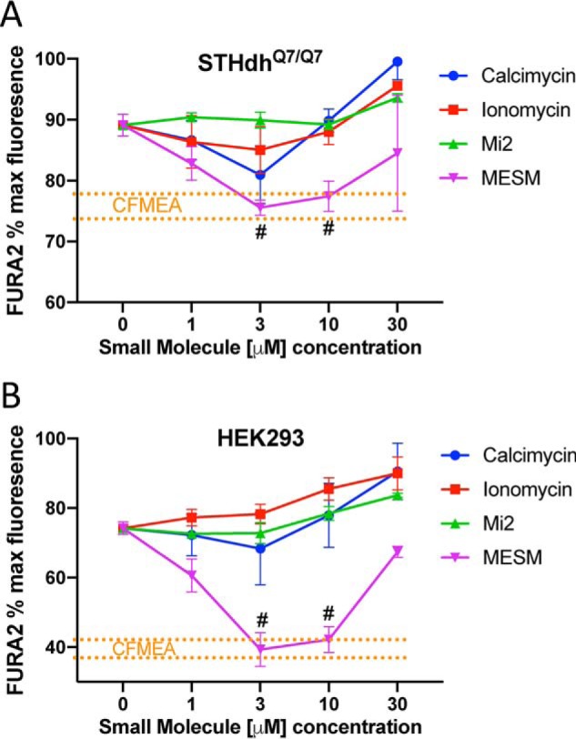
Figure 6.

Extraction with MESM is comparable with CFMEA output from 3 to 10 μm and outperforms other known Mn ionophores. Murine striatal neuron lineage of Q7 cells (A) or HEK cells (B) was exposed to 100 μm Mn for 2 h at 37 °C. Q7 cells (n = 3–6) were exposed to Mn in HBSS and HEK cells (n = 3) in DMEM, respectively. The cells were then washed five times in PBS (lacking Ca2+ and Mg2+) and then exposed to 0.5 μm Fura-2 and varying concentrations of calcimycin, ionomycin, manganese ionophore II (Mi2), or MESM for 15 min before reading at 360/535 nm excitation/emission. A separate group of cells had Mn extracted by traditional CFMEA means after Mn exposure, for comparison. CFMEA dotted lines (in orange) denote range of 1 S.D. from CFMEA average. Each point outside the orange lines is significantly different from CFMEA, as determined by a two-way ANOVA with Sidak's multiple comparisons. The points within the orange lines are not significantly different from CFMEA levels and are labeled with #. Error bars represent standard deviation of all biological replicates.
