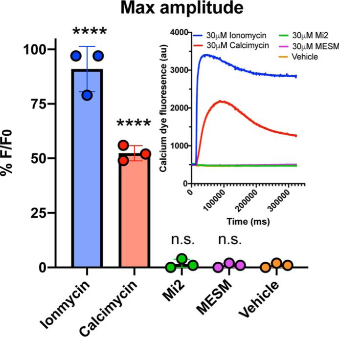
Figure 9.

MESM does not influence intracellular calcium. HEK293 cells (n = 3, each experiment was performed in triplicate wells) were plated the day before the experiment at 15,000 cells per well and grown until 100% confluency. Cells were soaked with Fluo-4 dye for 1 h at room temperature prior to adding varying concentrations of MESM, ionomycin, calcimycin, and manganese ionophore II (Mi2) ranging from 3 nm to 30 μm. Shown here is the maximum concentration (30 μm). Using PanOptic to measure fluorescence every second for 5 min, an ionophore from a donor plate was added to the acceptor plate containing cells. Fluorescence was measured at 482 ± 16 nm excitation and 536 ± 20 nm emission. Inset shows a representative trace of each ionophore at maximum concentration (30 μm). The maximum amplitude was calculated after normalizing for light variations with a static ratio and subtracting background. Percent fluorescence was normalized to the maximum fluoresced wells in each plate (30 μm ionomycin). An ordinary one-way ANOVA was performed with Dunnett's multiple comparisons test. Statistical significance from vehicle is indicated as not significant (n.s.) or **** (p < 0.0001).
