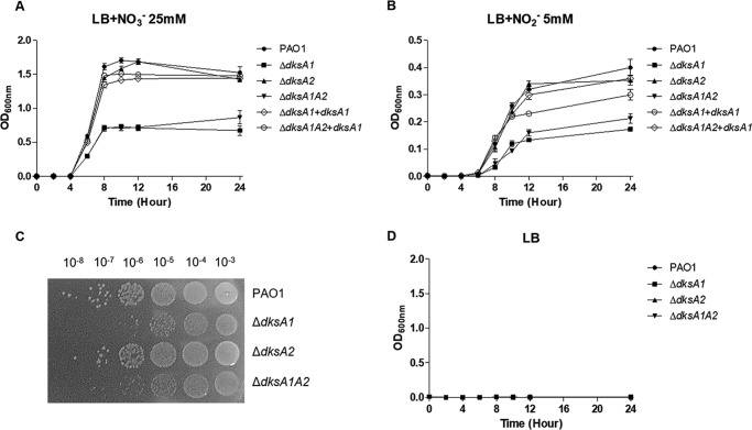
Figure 4.
Anerobic growth of ΔdksA1, ΔdksA2, and ΔdksA1ΔdksA2 mutants. A, growth curves of PAO1, ΔdksA1, ΔdksA2 and ΔdksA1ΔdksA2 strains in LB medium supplemented with 25 mm NO3−. Aliquots of bacterial cultures (n = 3) were withdrawn every 2 h to measure OD600 values. Bacterial strains (indicated to the right) were grown anaerobically at 37 °C. B, growth curves of PAO1, ΔdksA1, ΔdksA2, and ΔdksA1ΔdksA2 strains in LB medium supplemented with 5 mm NO2−. C, defective anaerobic growth of ΔdksA1 and ΔdksA1ΔdksA2 was further verified in cfu enumeration assay. Bacterial cultures after 24 h of anaerobic growth in LB supplemented with 25 mm NO3− were serially diluted, and 10 μl of each diluent was spot-inoculated onto an LB agar plate. The plates were incubated in anaerobic conditions at 37 °C for 24 h. D, anaerobic growth was not active in plain LB medium without either alternative electron acceptor. Error bars, S.D.

