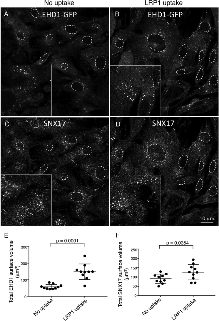
Figure 3.
EHD1 is recruited to endosomes upon LRP1 uptake. A–D, CRISPR/Cas9 gene-edited NIH3T3 cells expressing EHD1-GFP were either mock-treated (A and C, no uptake) or incubated with anti-LRP1 antibody (B and D, 30 min on ice and 30 min at 37 °C) prior to fixation and immunostaining with anti-SNX17 antibody and imaging by confocal microscopy. Representative images consisting of a field of cells are displayed. Regions of interest are shown in the insets, and dashed ovals outline the nuclei of the cells. E, 3D surface rendering was carried out from z-sections to capture and quantify the total surface volume of EHD1 (E) or SNX17 (F) (see “Experimental procedures” for details). Error bars denote standard deviation. Two-tailed t tests were performed to derive p values. Data shown are representative of three independent experiments, each using 10 images with seven z-sections each.

