Figure 1.

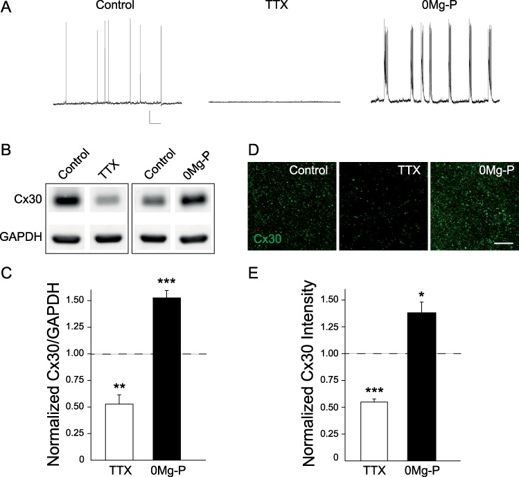
Activity-dependent regulation of Cx30 protein levels. (A) Spontaneous activity of hippocampal CA1 pyramidal cells recorded in current clamp in control, TTX (0.5 μM, 1–3 h) and 0Mg-P (100 μM, 1–3 h) conditions. Scale bar, 20 mV; 6.7 s. (B) Immunoblot detection of Cx30 in hippocampal acute slices in regular ACSF (control), ACSF containing TTX (0.5 μM, 1 h), or 0Mg-P (100 μM, 3 h). GAPDH was used as a loading control. (C) Quantitative analysis of Cx30 protein levels showing that Cx30 expression was reduced in TTX (n = 7) and increased in 0Mg-P (n = 17). Relative expression levels of Cx30 normalized to GAPDH levels; control ratio is set to 1. (D) Immunofluorescent staining for Cx30 in the hippocampal CA1 region from acute slices in regular ACSF (control), ACSF containing TTX (0.5 μM, 1 h) or 0Mg-P (100 μM, 3 h). Scale bar, 25 μm. (E) Quantification of Cx30 staining intensity; control intensity level is set to 1. Cx30 immunostaining intensity was decreased in TTX (n = 9) and increased in 0Mg-P (n = 9) compared to control ACSF (n = 9; ANOVA and Dunnett’s post hoc test). Asterisks indicate statistical significance (*P < 0.05, **P < 0.01, ***P < 0.001).
