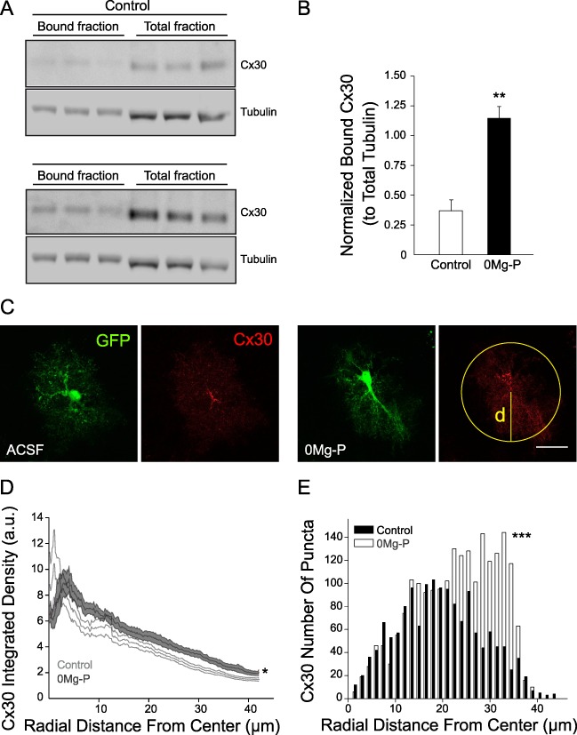
Figure 4.

Cx30 subcellular localization is regulated by neuronal activity. (A) Activity-dependent Cx30 plasma membrane trafficking measured by hippocampal acute slices biotinylation. Immunoblot detection of total and surface Cx30 proteins in slices incubated in ACSF or 0Mg-P (100 μM, 3 h). Tubulin was used as a loading control. (B) Quantitative analysis of normalized surface Cx30 protein levels (n = 3). (C) Immunofluorescent staining for Cx30 (red) within eGFP-expressing hippocampal CA1 astrocytes (green) from acute slices incubated in ACSF or 0Mg-P (100 μM, 3 h). Cx30 intensity profile was determined at any given radial position as the sum of the pixel values around a circle centered at the cell soma, as shown in yellow. (D) Quantification of Cx30 radial intensity profile (n = 9 cells). Cx30 intensity was increased in distal astrocytes processes (~ 20–40 μm from the center of the cell soma) in 0Mg-P condition. Scale bar, 25 μm. Asterisks indicate statistical significance (*P < 0.05, **P < 0.01).
