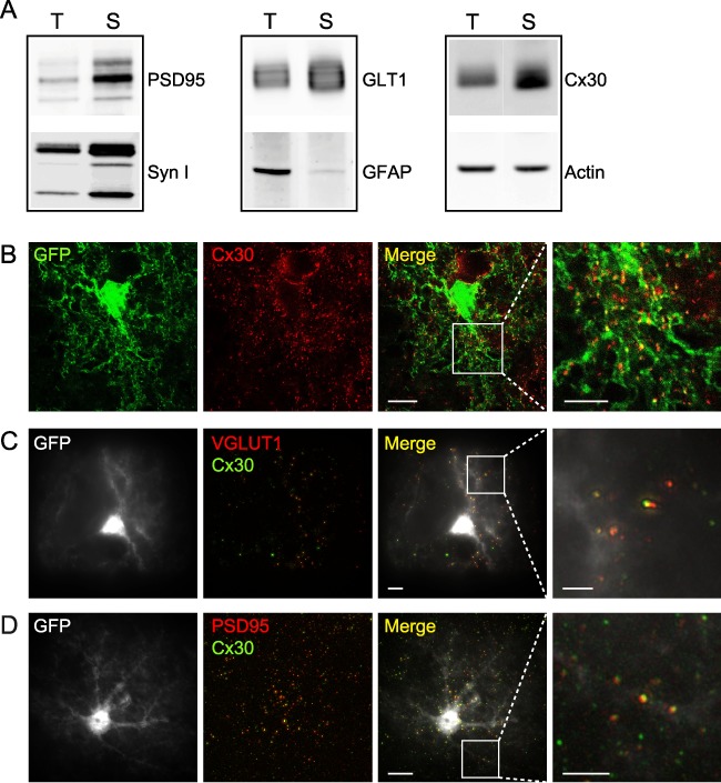
Figure 5.

Cx30 is localized in distal perisynaptic astrocyte processes. (A) Western blotting detection of pre- (syn I) and post- (PSD95) synaptic proteins showed an enrichment of plasma membrane synaptic proteins in crude synaptosomal membrane fraction (S) compared to total hippocampal fraction (T), while actin did not change. Astroglial GLT1 glutamate transporters are also enriched in synaptosomal fractions, while GFAP protein levels are very low. Astroglial Cx30 is strongly expressed within crude synaptosomal membrane fraction (n = 6). (B) Confocal/STED images of GFAP-eGFP (green) mouse brain slices immunostained for Cx30 (red). The stack merged image reveals the localization of Cx30 STED-resolved puncta (ATTO 647, 1:250) within distal GFP-labeled astrocyte processes. Scale bar, 10 μM. (C) SIM images of GFAP-eGFP (white) mouse brain slices immunostained for VGLUT1 (red) and Cx30 (green). The single-plane merge image indicates occasional colocalization of Cx30 with the presynaptic protein VGLUT1. Scale bar, 10 μM. (D) SIM imaging of GFAP-eGFP (white) mouse brain slices immunostained for PSD95 (red) and Cx30 (green). The single-plane merge image reveals occasional colocalization of Cx30 with the postsynaptic protein PSD95. Scale bar, 10 μM. White squares in (B–D) indicate the small regions of interest that are magnified. Scale bar, 5 μM.
