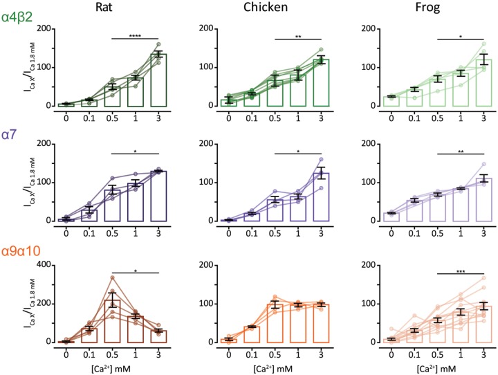
FIg. 6.
Extracellular Ca2+ potentiates neuronal nAChRs but differentially modulates α9α10 nAChRs. ACh response amplitude as a function of extracellular Ca2+ concentration. ACh was applied at near-EC50 concentrations (10 μM ACh for all α4β2, rat and chick α9α10 nAChRs and 100 μM ACh for all α7 and frog α9α10 nAChRs). Current amplitudes recorded at different Ca2+ concentrations in each oocyte were normalized to the response obtained at 1.8 mM Ca2+ in the same oocyte. Vh: −90 mV. Bars represent mean ± S.E.M., open circles represent individual oocytes (n = 4–12). *P < 0.05, **P < 0.01, ***P < 0.005, and ****P < 0.0001, paired t-test (rat and frog α4β2 nAChRs and all α7 and α9α10 nAChRs) or Wilcoxon matched pair test (chick α4β2 nAChR)—comparing 0.5 mM Ca2+ versus 3 mM Ca2+.

