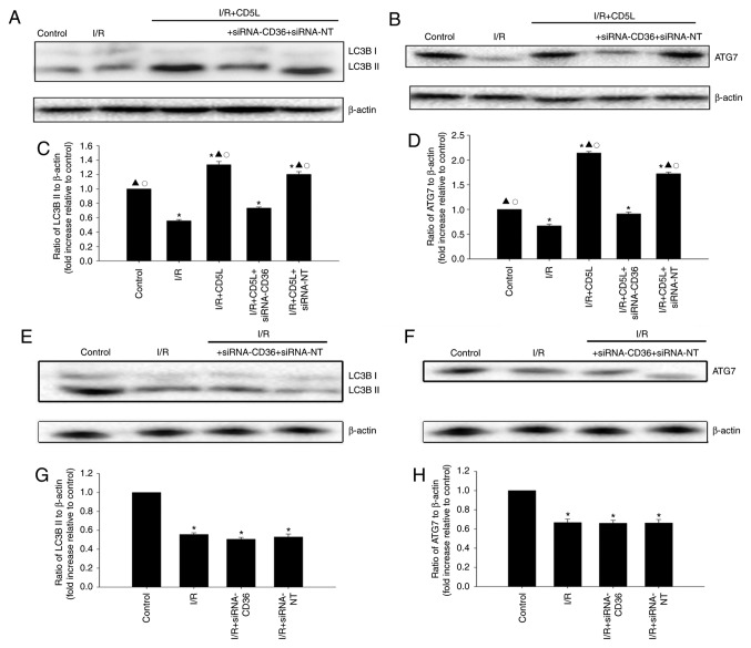
Figure 3.
CD5L activates the cellular autophagy process. (A-D) Hepatocytes were transfected with siRNA-CD36 or siRNA-NT, incubated under I/R conditions and treated with CD5L. Cells without any treatment was used as control. (A and C) Representative western blots and quantification of the expression levels of LC3B-II and β-actin. (B and D) Representative western blots and quantification of the expression levels of ATG7 and β-actin. Each column represents the mean ± SD of three independent experiments. *P<0.05 vs. control; ▲P<0.05 vs. I/R; ○P<0.05 vs. I/R + CD5L + siRNA-CD36. (E-H) Hepatocytes were transfected with siRNA targeting CD36 or siRNA-NT as a control and incubated under I/R conditions. (E and G) Representative western blots and quantification of the expression levels of ATG7. (F and H) Representative western blots and quantification of the expression levels of β-actin. Each column represents the mean ± SD of three independent experiments. *P<0.05 vs. control. I/R, ischemia/reperfusion; CD5L, CD5-like; CD36, cluster of differentiation 36; siRNA, small interfering RNA; NT, non-targeting.

