Figure 1.
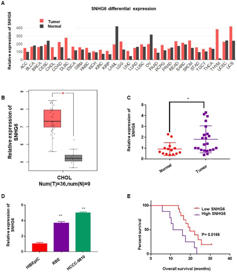
SNHG6 was overexpressed in CCA tissues and cells, and associated with poor overall survival in CCA patients. (A) SNHG6 expression (TPM) in different human malignancies and their matched normal tissues from the GEPIA software (http://firebrowse.org). (B) SNHG6 expression (Log2(TPM+1) in CCA tissues (n=36) compared with noncancerous tissues (n=9) from TCGA database analyzed with GEPIA software. (C) The relative expression (normalized to β-actin) of SNHG6 in CCA tissues (n=22) and adjacent normal tissues (n=14). (D) Relative SNHG6 expression in two CCA cell lines (HCCC-9810 and RBE) and one normal biliary epithelial cell line HIBEpiC. (E) Kaplan-Meier survival plots showed that high SNHG6 expression was correlated with poor OS in CCA patients (n=22, p=0.0166).

