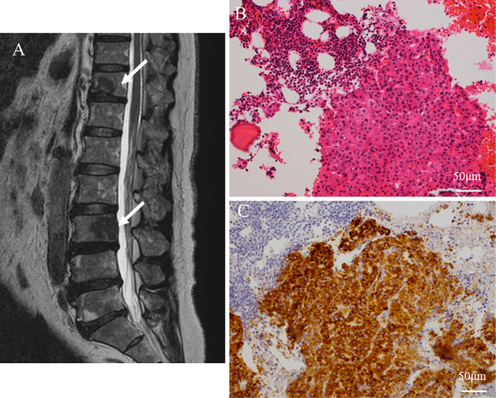
Figure 2.
Bone metastases of hepatocellular carcinoma. Magnetic resonance imaging revealed metastatic bone tumors at the Th-11 and L-3 vertebrae (arrows) (A), and then a bone biopsy was performed at the L-3 vertebra. Histopathological examinations of the bone biopsy specimens showed a thick trabecular pattern of tumor cells (Hematoxylin and Eosin staining) (B). Immunohistochemical staining was positive for Hep Par 1, suggesting metastatic hepatocellular carcinoma (Hep Par 1) (C).

