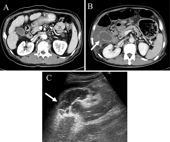
Figure 4.
Imaging of the first gallbladder perforation. CT revealed no gallbladder stones or gallbladder metastases of HCC (A). CT showed rupture of the gallbladder wall (arrow) and ascites around the gallbladder, suggesting gallbladder perforation (B). US also indicated rupture of the gallbladder wall (arrow) (C). CT: computed tomography, HCC: hepatocellular carcinoma, US: ultrasonography

