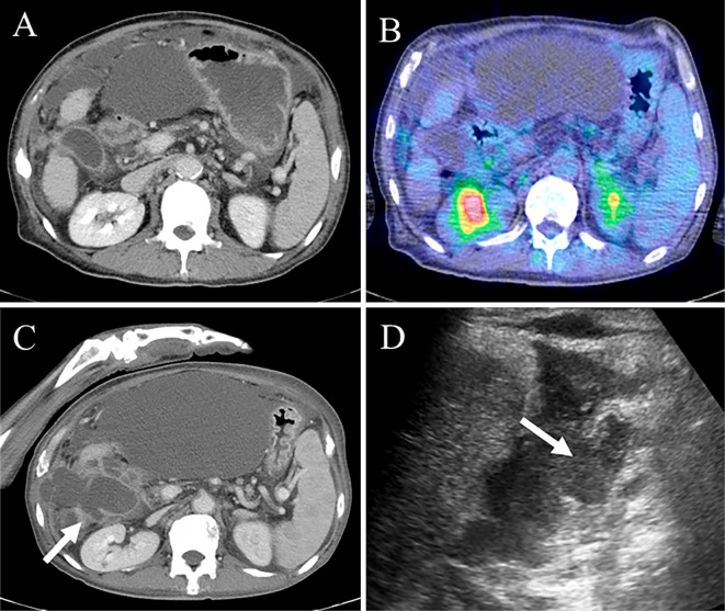
Figure 5.
Imaging of the second gallbladder perforation. CT showed improvement in the gallbladder perforation, but massive ascites remained (A). The 18F-FDG PET/CT findings did not show any FDG uptake in the gallbladder (B). CT and US showed rupture of the gallbladder wall (arrow) (C, D). CT: computed tomography, 18F-FDG PET/CT: 18F-fluorodeoxy glucose-positron emission tomography/computed tomography, US: ultrasonography

