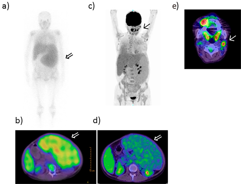
Figure 2.
Gallium and PET-CT scans. a, b: Gallium scan and c-e: PET-CT scan. Gallium scans showed accumulation in the spleen (arrow ⇒) (a, b). The neck lymph node was positive (arrow ↑) (c, e); however, massive splenomegaly was negative (arrow ⇒) (d) on PET-CT. PET-CT: positron emission tomography-computed tomography

