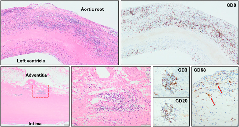
Figure 7.
Histopathological findings (Hematoxylin and Eosin staining and immunostaining). Predominant and diffuse infiltration of CD8+ T-lymphocytes into the fibrosa of the aortic valvar leaflet (upper panels) is observed. On the adventitia of the sinuses of Valsalva (lower panels), CD8+ T-lymphocytes, CD20+ B-lymphocytes, and CD68+ multinucleated giant cells (red arrows) infiltrated predominantly around the vasa vasorum. The lower middle panel shows a magnified view of the red dashed box in the lower left panel.

