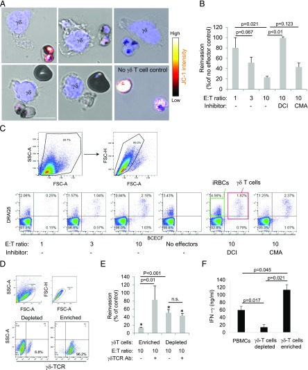
FIGURE 3.
γδ T cells reduce P. falciparum viability in iRBCs and the reinvasion capacity of the parasites. (A) Activated γδ T cells were incubated with JC-1–prestained late-stage iRBCs for 30 min and then stained with Hoechst as nuclear marker (blue) before JC-1 staining intensity was assessed by life cell microscopy. Scale bar, 10 μm. (B) Activated γδ T cells, prelabeled with BCECF-AM, were incubated with late-stage iRBCs for 2 h at indicated E:T ratios before dilution in uninfected RBCs to assess reinvasion capacity. After 48 h, the cultures were stained with DRAQ5, and parasitemia was measured by flow cytometry. DCI and CMA were used to inhibit Gzm and PFN activity, respectively. (C) The gating strategy of a representative experiment is presented. (D) We show a representative flow cytometry analysis using γδ TCR Abs of cells that were MACS purified (enriched for or depleted of γδ T cells) and further activated with iRBC supernatant and IL-2 for 6 d. (E) These purified cells (γδ-enriched versus γδ-depleted PBMCs) were tested in reinvasion assays as above, including the addition of a blocking γδ TCR Ab to the effector cells just before the coincubation with iRBCs. In addition, the supernatant of enriched γδ T cells compared with γδ T cell–depleted cultures or unpurified PBMCs after 10 d of activation with iRBC supernatant and IL-2 were tested for INF-γ levels by ELISA. Averages ±SEM of the normalized reinvasion capacity of four independent experiments are shown (B, E, and F). The p values of differences between groups, calculated by Student t test, are indicated. (E) The asterisks (*) represent statistical differences (Student t test) to no-effector cell controls.

