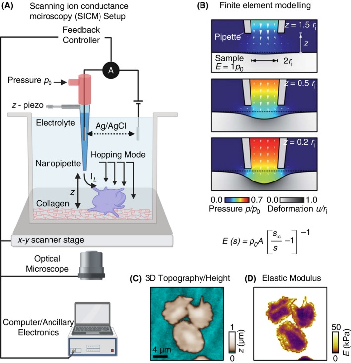
Figure 4.

A, Schematics of scanning ion conductance microscopy (SICM) used for high‐resolution topography imaging and biomechanical characterization of adherent platelets. The SICM setup consists of a pressure‐controlled borosilicate glass nanopipette with a Ag/AgCl electrode and a second electrode connects the electrolyte‐filled culture dish with the nanopipette. The culture dish is mounted on a sample scanner that drive in x and y‐axis (for lateral movement) and z‐piezo motors (for vertical scanning). An applied voltage between 2 electrodes induces an ionic leakage current IL through the electrolyte‐filled nanopipette, which depends on the distance d between pipette and sample. A controller records this ion current and drives the xy‐ and z‐piezo. For cell biological applications the SICM can be easily integrated with an optical microscope. B, Theoretical modeling using in silico finite element mechanics (FEM) simulation and calculations showing resulting deformation of an elastic sample as a function of the vertical SICM nanopipette position upon application of fluid flow induced by the pressure p0 applied to the upper pipette end.; C, Representative SICM 3D topography and Young’s modulus mapping of spreading platelets. (Figure adapted and modified from Rheinlaender and Schaffer65 and Rheinlaender et al.66)
