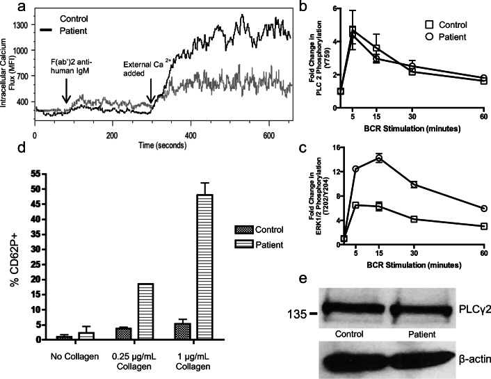
Fig. 4.
Evidence of altered BCR signaling. a Calcium flux in primary CD19+ B cells after BCR stimulation. Fresh PBMCs from patient (PLCγ2M1141K) and healthy control were incubated in Ca2+-free media and stimulated via BCR followed by addition of exogenous Ca2+. Representative of three independent blood collections including one prior to initiation of IVIG therapy with three different sex-matched pediatric controls. b, c ERK1/2 phosphorylation after BCR stimulation. Primary CD19+ B cells from patient (PLCγ2M1141K) and healthy control were stimulated for the indicated times and the phosphorylation of PLCγ2 (b) and ERK1/2 (c) was measured by intracellular flow cytometry. These experiments were repeated with three blood draws from the patient. Each data point is presented as a ratio of MFI to baseline MFI at start of incubation. Values represent means ± SEM. d Collagen-induced platelet alpha granule release. Fresh blood was collected without use of a tourniquet from the patient (PLCγ2M1141K) and healthy controls into sodium citrate tubes with a discard tube first and stimulated with collagen. Alpha granule release from platelets was assessed by measuring the surface expression of CD62P on CD61+ platelets by flow cytometry. Each column represents data from 3 individual samples. e Equal amounts of PLCγ2 protein in about 1 million isolated CD19+ B cells was confirmed by immunoblotting. Representative of two independent collections

