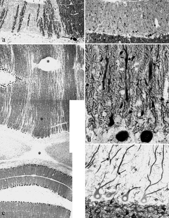
Fig. 1a–e.

Purkinje cell dendrites in cat 2 ( a, b, d) and cat 3 ( c, e). Anti-calbindin ( a, c, d) and anti-MAP2 ( b, e). a, b Corresponding area from two adjacent transversal sections showing gaps in calbindin-positive dendrites in the molecular layer ( a) with normal numbers of stellate and basket cells ( b). One Purkinje cell body is visualized by both immunesera (arrows in a and b ). A blood vessel (v) is also visible in both sections. c Symmetrical parasagittal gaps without calbindin immunoreactivity in the vermis. The nodulus (bottom of the figure) is spared. The stars indicate the midline. d Dendritic swellings (arrows) and abnormal branching. e Empty vacuoles (arrows). Bar = a, b:160 µm; c:320 µm; d:40 µm; e:60 µm
