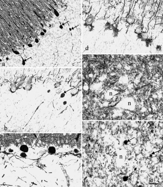
Fig. 2a–f.

In the granular layer ( a – d) of cats 2 ( a) and 3 ( b, c, d), numerous spheroids are present on the Purkinje cell axons, stained by anti-calbindin (arrows in a ) and by clone RT97 (arrow in b ). Moderately increased calbindin-positive recurrent synapses are visible in c. RT97-positive baskets are either normal ( b) or hypertrophied ( d). Neurons (n) in the normal deep cerebellar nuclei of cat 1 ( e) are covered by calbindin-positive synapses, which are almost absent in cat 3 ( f). The arrows in f point out spheroids. Bar = a:83 µm; b:30 µm; c, d:35 µm; e, f:22 µm
