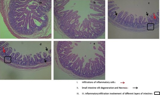
Fig. 5.

Representative images of histological changes in the small intestine from piglets fed sera from pigs immunized with (a) rSPV-St, (b) inactivated SQ2014, (c) wtSPV, or (d) PBS, followed by challenge with PEDV SQ2014. The samples were obtained 3 days post-challenge. The intestinal tissue from a normal healthy pig (e) was included for comparison. The tissues were stained with hematoxylin and eosin (HE). Magnification, 100×
