
Fig. 2.
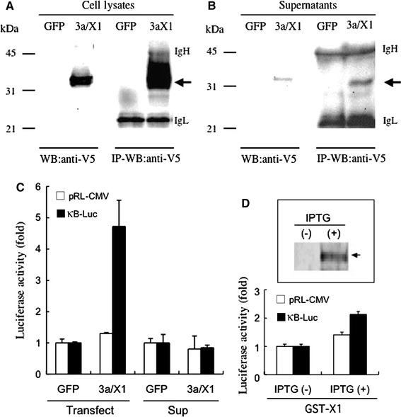
Secretion of SARS-CoV 3a/X1 from cells. (a, b) Cell lysates (a) and culture supernatants (b) from HEK293T cells that had been transfected with pLenti/V5/GFP (GFP) or pLenti/V5/X1 (3a/X1) 48 h earlier were subjected to Western blot (WB) analysis with anti-V5 antibody directly (WB) or following immunoprecipitation with the same antibody (IP-WB). Arrows indicate the sizes of 3a/X1. Sizes of immunoglobulin heavy (IgH) and light chains (IgL) are also indicated. (c) Effects on NF-κB activity of direct expression of 3a/X1 (Transfect) and soluble 3a/X1 (Sup). For the direct expression experiment, HEK293T cells were co-transfected with NF-κB reporter plasmid (κB-Luc) (closed bar), pRL-CMV (open bar), and pLenti/V5/GFP (GFP) or pLenti/V5/X1 (3a/X1), followed by 48 h culture (Transfect). For the soluble-3a/X1 experiment, κB-Luc (closed bar)- or pRL-CMV (open bar)-transfected HEK293T cells were cultured for 24 h in the culture supernatants of separately prepared HEK293T cells that had been transfected with pLenti/V5/GFP (GFP) or pLenti/V5/X1 (3a/X1) 48 h earlier (Sup). Luciferase activities in cell lysates were indicated as fold increases against GFP controls. (d) Crude GST-3a/X1 fusion proteins were extracted from E. coli transformed with pGEX-X1 as described in Materials and methods. GST-3a/X1 fusion proteins in the E. coli extracts with or without IPTG treatment were detected by Western blot using antibodies to GST (top panel). Arrows indicate the sizes of GST-3a/X1 fusion protein (63 kD). These extracts (50 μl/well) were added to separately prepared HEK293T cells that had been transfected with κB-Luc (closed bar) and pRL-CMV (open bar) plasmids the day before. Luciferase activities in cell lysates were measured after a 24-h incubation. The data represent fold increases against the control sample incubated with E. coli extracts without IPTG induction, and indicated as the mean ± SD of duplicate samples