
Fig. 3.
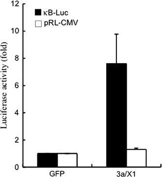
Activation of NF-κB by SARS-CoV 3a/X1 in RAW264.7cells. The NF-κB reporter plasmid (κB-Luc) (closed bar) and pRL-CMV (open bar) were introduced together with pLenti/V5/X1 (3a/X1) or control pLenti/V5/GFP (GFP) vectors into RAW264.7cells by transfection, and luciferase activities were measured approximately 48 h after transfection. Data represent means ± S.D. of fold increases against GFP controls in three independent experiments