
Fig. 4.
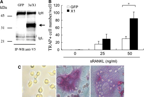
Augmentation of osteoclast differentiation by SARS-CoV 3a/X1. a RAW264.7 cells were infected with pseudotyped viruses expressing 3a/X1 (VSV-G/pLenti/V5/X1) or GFP (VSV-G/pLenti/V5/GFP), and the cell lysates 48 h after infection were subjected to Western blot analysis with anti-V5 antibody after immunoprecipitation with the same antibody. Arrows indicate the size of 3a/X1. b RAW264.7 cells were infected with VSV-G/pLenti/V5/GFP (open bar) or VSV-G/pLenti/V5/X1 (closed bar) pseudotyped viruses for 48 h, and 1,000 cells/spot were further cultured for 5 days with sRANKL at the indicated concentrations. The cells were stained with TRAP, and the osteoclast-like TRAP+ multinuclear cells in each well were counted under a microscope. Data are expressed as means ± SD of triplicate wells. *p < 0.05. c Representative images after TRAP staining of untreated RAW264.7 cells (left) and VSV-G/pLenti/V5/GFP-infected (middle) or VSV-G/pLenti/V5/X1-infected (right) RAW264.7 cells in the presence of 50 ng/ml sRANKL