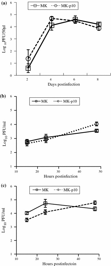
Fig. 3.

The replication kinetics of MK (squares) and MK-p10 (circles). a Viral replication in suckling mouse brain. Zero-day-old mice were infected i.c. with 5,000 PFU of PEDV. At the indicated days p.i., the brains were collected and 10% homogenates were prepared. The viral titer in the brains was determined using a plaque assay with Vero cells and is expressed as PFU/50 μl of 10% homogenates (n = 3). b, c Viral replication in cultured cells. Vero cells were infected with PEDV at an m.o.i. of 0.1. Viral titers in the supernatants (b) and cells (c) were determined separately using plaque assays (n = 6). The viral titer in the supernatant is given as PFU/ml of supernatant, while that in the cells is given as PFU per well of a 24-well plate
