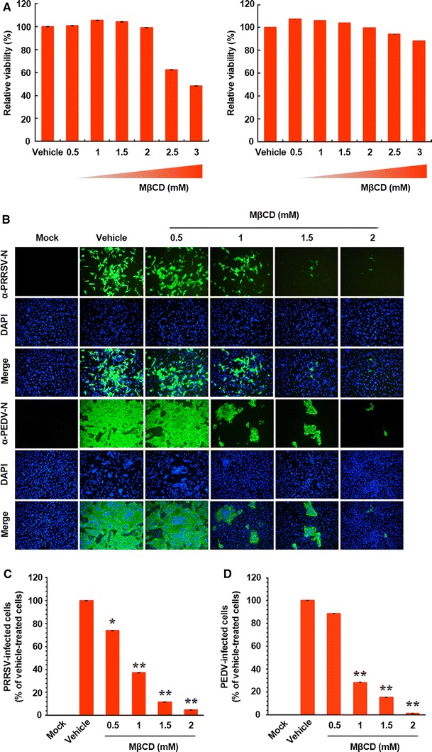
Fig. 1.

Effects of cellular cholesterol depletion on the replication of porcine nidoviruses. (A) PAM-pCD163 (left) and Vero (right) cells were incubated with various concentrations of MβCD for 48 h prior to the MTT assay, and the cytotoxicity of MβCD was determined by the MTT assay. (B) PAM-pCD163 and Vero cells were preincubated with MβCD at the indicated concentrations for 1 h prior to infection and were mock infected or infected with PRRSV or PEDV at an MOI of 1. Virus-infected cells were further maintained for 48 h in the presence of vehicle or MβCD. For immunostaining, infected cells were fixed at 48 hpi and incubated with MAb against the N protein of PRRSV or PEDV, followed by incubation with Alexa-green-conjugated goat anti-mouse secondary antibody (first and fourth panels). The cells were then counterstained with DAPI (second and fifth panels) and examined using a fluorescent microscope at 200× magnification. (C and D) Viral production in the presence of MβCD was calculated by measuring the percentage of cells expressing N proteins of PRRSV (C) or PEDV (D) by flow cytometry. The values shown are the means of three independent experiments, and error bars represent standard deviations. *, P = 0.001 to 0.05; **, P < 0.001
