
Fig. 2.
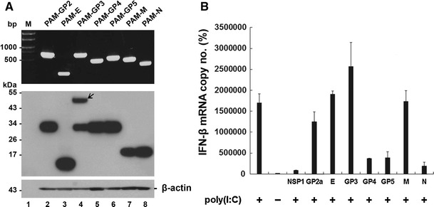
Involvement of specific PRRSV structural proteins in inhibition of IFN-β induction. (A) Established stable PAM cell lines were grown independently for 48 h, and total RNA was extracted from cells. Each viral gene was amplified by RT-PCR and visualized on a 0.8% agarose gel (top panel). Cell lysates were prepared from cells and subjected to western blot using an anti-His tag antibody to determine the expression level of each PRRSV protein in stable cell lines (middle panel). The blot was also reacted with anti-β-actin antibody to confirm equal protein loading (bottom panel). Lane 1 (M), molecular weight marker; lane 2, PAM-GP2; lane 3, PAM-E, lane 4, PAM-GP3, lane 5, PAM-GP4; lane 6, PAM-GP5; lane 7, PAM-M; lane 8, PAM-N. (B) PAM cells stably expressing each structural protein of PRRSV were stimulated with poly (I:C) as described in “Materials and methods”. Total RNA isolated from cells was subjected to real-time RT-PCR, and the porcine IFN-β mRNA levels in each stable cell line were expressed as copy numbers relative to the unstimulated control. These data are representative of the mean values from three independent experiments, and error bars represent standard deviations