
Fig. 5.
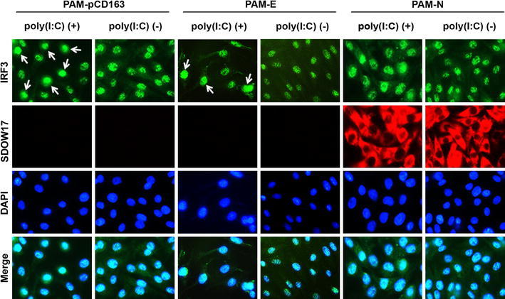
Subcellular localization of IRF3 in the presence of N. PAM-pCD163 and PAM cells stably expressing N or E were grown for two days and stimulated with poly (I:C) for 6 h. Cells were fixed with 4% paraformaldehyde and co-stained with anti-IRF3 and PRRSV N or His-tag antibodies, followed by incubation with a mixture of Alexa Fluor 488 (green)-conjugated goat anti-rabbit and 594 (red)-conjugated goat anti-mouse secondary antibodies (top and second panels). The cells were then counterstained with DAPI (third panels) and examined using a fluorescent microscope at 400× magnification. Arrows indicate nuclear localization of IRF3 upon poly(I:C) stimulation (color figure online)