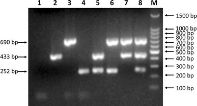
Fig. 1.

Agarose gel electrophoresis (1.0%) of specific fragments amplified by mPCR from proviral DNA and cDNAs of specific CPV-2, CAV, and CCoV isolates. Lane 1, negative control; lane 2, CAV (433-bp fragment); lane 3, CPV-2 (690-bp fragment); lane 4, CCoV (252-bp fragment); lane 5, CCoV/CAV; lane 6, CPV-2/CCoV; lane 7, CAV/CPV-2; lane 8, CPV-2/CAV/CCoV; lane M, 100-bp DNA ladder marker
