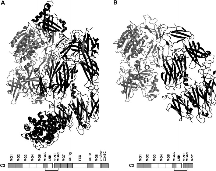
Fig. 1.

Structural implications for the p.Val899AlafsX5 mutation. The 3D structure of the active C3 cleavage product C3b (black) in complex with CFB (gray) and the domain structure of C3 are depicted for both the wild-type (a) and the mutant protein (b). In gray, the C3 domains involved in CFB binding are indicated [5]. The images were generated using PyMOL with the Protein Data Bank file 2XWB
