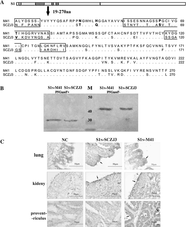
Fig. 1.

(A) Amino acid sequence alignment of S1N (residues 19–270) of M41 and SCZJ3. Four main divergent regions are located at aa 20-26, 53-80, 117-122, 129-136. The critical amino acids for M41 (N38T, H43Q, P63S, T69V) are shown in bold. (B) Western blot. S1N-SCZJ3 and S1N-M41 were purified using an Ni-NTA column and analyzed by Western blot. The samples were both recognized by polyclonal IBV-M41 antiserum before and after treatment with PNGase F. (C) Histochemistry was performed by incubating S1N-M41 and S1N-SCZJ3 with lung, kidney and proventriculus samples of PBS as control. Binding to peribronchial epithelial cells in lung tissue and to the renal tubular epithelial cells in kidney were detected. Distinctive binding of S1N-SCZJ3 to mucous membrane epithelial cells was detected in the proventriculus
