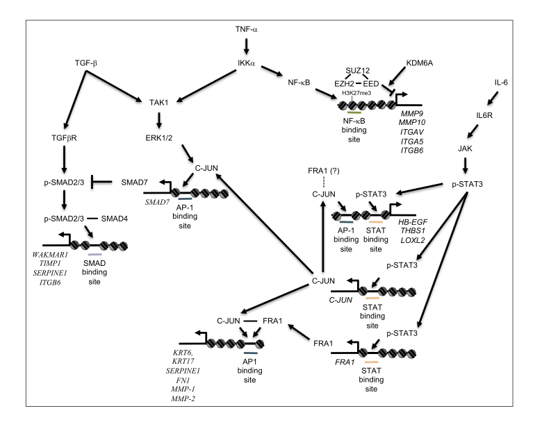
Figure 2.
Transcriptional network regulating initiation of re-epithelialization. Schematic diagram of signaling pathways activated by cytokines and growth factors present in the wound bed, leading to initiation of re-epithelialization. TGF-β, TNF-α, and IL-6 released into the wound environment activate interconnected pathways, which collaborate with AP-1 transcription factors expressed in wound edge keratinocytes to activate transcription of target genes (Italics) required for matrix re-modeling and initiation of keratinocyte migration.

