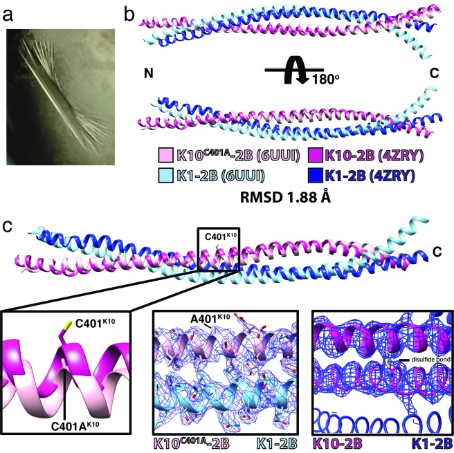
Figure 4.
Human K1/K10C401A-2B heterodimer crystal structure. (a) Representative crystal of the K1/K10C401A-2B complex. (b) Superposition of the K1/K10C401A-2B heterodimer onto the wild-type K1/K10-2B heterodimer (PDB ID 4ZRY) had a root-mean-square deviation (RMSD) of 1.88 Å across all atom pairs. There is enhanced curvature at the C-terminus of K1-2B in the K1/K10C401A-2B heterodimer (light blue) compared to the wild-type structure. (c) A zoomed in view of the superposed structures from (b) at K10 position 401 demonstrated electron density for an alanine, confirming the K10C401A substitution. There is no structural evidence for a disulfide bond in the K1/K10C401A-2B heterodimer.

