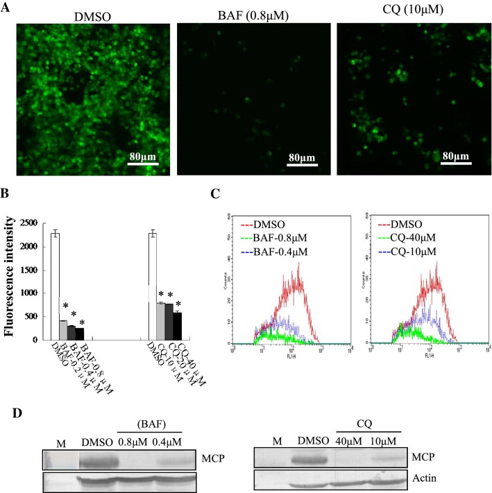
Fig. 5.
STIV enters into cells in a pH-dependent manner. (A) Fluorescence microscopy of EGFP-STIV- infected cells after treatment with DMSO, BAF or CQ. (B) Fluorescence intensity of EGFP-STIV-infected cells after treatment with DMSO, BAF or CQ. (C) Quantitative analysis of the percentage of EGFP-STIV-infected cells by flow cytometry. (D) Western blotting detection of protein synthesis of the STIV major capsid protein after treatment with BAF or CQ

