
Fig. 1.
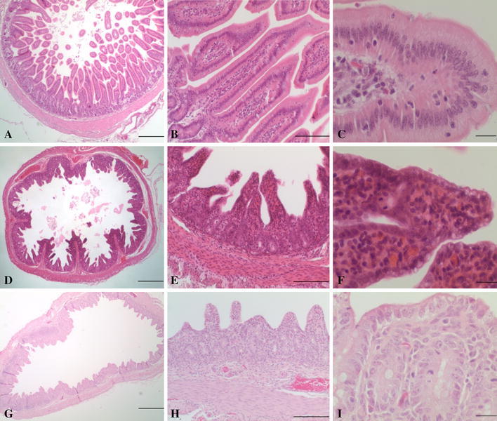
Photomicrographs depicting jejunum sections from nursing piglets not infected with a coronavirus (A-C), infected with porcine deltacoronavirus (PDCoV) (D-F), and infected with porcine epidemic diarrhea virus (PEDV) (G-I). A, jejunum; note villi of normal length (villus:crypt ratios >3-4:1). Bar = 500 µm. B, jejunum; villi are of normal length and are lined by tall columnar superficial enterocytes. Bar = 100 µm. C, jejunum; high magnification of superficial enterocytes with abundant pale eosinophilic cytoplasm. Bar = 20 µm. D, jejunum; moderate blunting of villi increasing the diameter of clear luminal space. Bar = 500 µm. E, jejunum; note decreased villus:crypt ratios (between 2:1-3:1) with fusion of two villi in the center of the image. Small amounts of cellular debris are evident in the lumen of scattered crypts. Bar = 100 µm. F, jejunum; superficial enterocytes have cuboidal morphology, and the cytoplasm is often vacuolated. Note villous fusion as well as a mitotic figure in the crypt epithelial cell. Bar = 20 µm. G, jejunum; severe diffuse villous atrophy with frequent fusion of villi. Bar = 500 µm. H, jejunum; marked villous atrophy with villus:crypt ratios of 1:1-2:1. Marked cuboidal attenuation of superficial enterocytes is present. Bar = 100 µm. I, jejunum; cytoplasmic vacuolation of attenuated superficial enterocytes is shown, as are mitotic figures in crypt epithelial cells. Note the collapse of the lamina propria stroma. Bar = 20 µm