
Fig. 1.
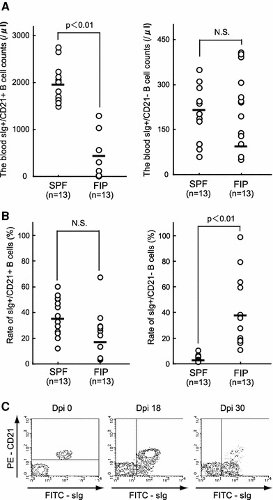
Emergence of sIg+ CD21− B-cells after FIPV infection and their counts and ratio in PBMCs: PBMCs were isolated from SPF and FIP cats, and B-cell surface antigens (sIg and CD21 molecules) were analyzed by flow cytometry. a The counts of sIg+ CD21+ B-cells and sIg+ CD21− B-cells in PBMCs, b the ratio of sIg+ CD21+ B-cells and sIg+ CD21− B-cells in PBMCs, c sIg and CD21 antigen expression on the feline B-cell surface upon FIPV infection (Dpi 0), day 18 after FIPV infection (Dpi 18), and the FIP onset time (Dpi 30). N.S. not significant, Dpi day post-inoculation