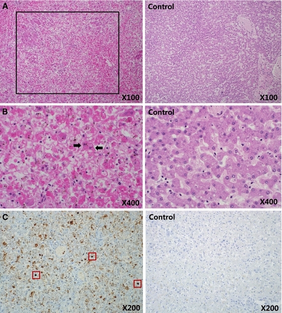
Fig. 1.

Massive hepatic necrosis (black box) (A, ×100). Basophilic intranuclear inclusion bodies (arrows) were observed in degenerated hepatic cells (B, ×400). The nucleic acid of MVC (dark brown staining, red boxes) was detected in liver tissue (C, ×200) via in situ hybridization (color figure online)
