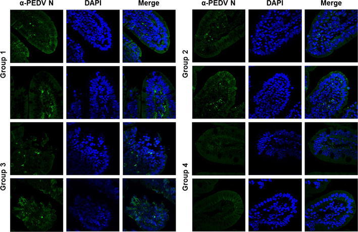
Fig. 6.

Detection of PEDV in small intestine tissues of piglets. Tissue specimens were prepared from small intestine of piglets from each group at the time of necropsy. The formalin-fixed and paraffin-embedded tissue sections were deparaffinized and subjected to immunofluorescence staining with an anti-PEDV N antibody. The sections were then counterstained with DAPI and examined using a fluorescence microscope at 200× magnification
