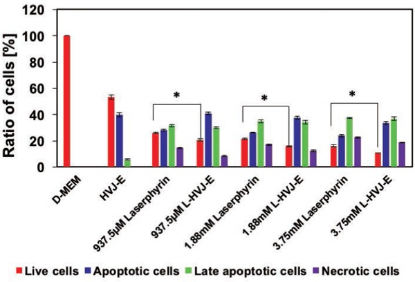
Figure 10:

Ratio of live, apoptotic, late apoptotic, and necrotic cells after PDT (quantification of fluorescence microscopic images shown in Fig. 9). Cell death type was assessed by annexin V–FITC and PI 24 h after PDT. The proportion of live cells was lower in L-HVJ-E–treated samples than in Laserphyrin®-treated samples, while the proportion of apoptotic cells was higher in L-HVJ-E–treated samples. Necrotic cells were more abundant in samples treated with 3.75 mM Laserphyrin® than with lower doses (1.88 mM or 937.5 µM), indicating that a higher photosensitizer concentration was more likely to induce necrosis than apoptosis.
