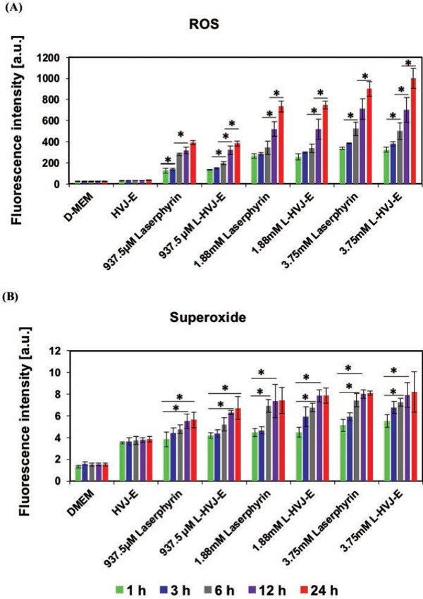
Figure 3:

Relative levels of (A) ROS and (B) superoxide produced in PC-3 cells following PDT. PC-3 cells were incubated in D-MEM, HVJ-E suspension, Laserphyrin® solution, or L-HVJ-E suspension at various Laserphyrin® concentrations for various incubation periods, followed by light irradiation at a wavelength of 664 nm (9 J/cm2). After PDT, the samples were immediately stained using the ROS-ID® total ROS/superoxide detection kit, and fluorescence intensities were measured using a fluorescence microplate reader. The highest levels of ROS and superoxide, produced after incubation for 24 h in L-HVJ-E suspension prepared with 3.75 mM Laserphyrin®, were ∼9-fold higher than in the D-MEM group. Both intracellular ROS and superoxide levels were significantly elevated by Laserphyrin® and L-HVJ-E-mediated PDT in a Laserphyrin® concentration and incubation period–dependent manner.
