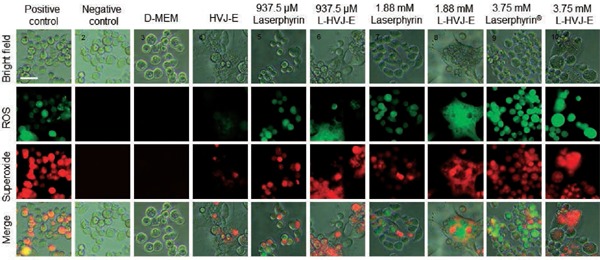
Figure 4:

Fluorescence microscope images of ROS and superoxide produced in PC-3 cells by Laserphyrin® and L-HVJ-E-mediated PDT. PC-3 cells were incubated in D-MEM, HVJ-E suspension, Laserphyrin® solutions, or L-HVJ-E suspensions for 3 h, followed by light irradiation at a wavelength of 664 nm (9 J/cm2). After PDT, the cells were stained with ROS-ID® total ROS/superoxide detection kit and observed under a fluorescence microscope. A small amount of ROS and superoxide was produced in the sample treated with HVJ-E, whereas no ROS/superoxide production was observed in the control sample. The highest levels of ROS and superoxide were detected in the sample treated with 3.75 mM L-HVJ-E suspension. Photomicrographs of positive and negative controls were obtained to ensure that the kit functioned as expected.
